THE BASIC PRINCIPLES OF COMPUTED RADIOGRAPHY
By : Sumarsono
Computed radiography (also known as diagnostic digital radiography), often abbreviated CR, refers to conventional projection radiography, in which the image is acquired in digital format using an imaging plate rather than film. Computed radiography (CR) system was introducing the image plate (IP), a product of the latest technology which has made a great, in detecting and recording X-ray images information of high quality and sensitifity. CR image can also be viewed on workstation. The images will be quickly transmitted through electrical lines and display on Cathode Ray Tube (CRT). Image processing using CRT will assist radiologist in making high quality diagnosis. Furthermore, storing images on optical disk will facilitate efficient archieving, even when space is limited.
The Computed Radiography System
Computed radiography is performed by a system consisting of the following functional components:
• A digital image receptor (The Imaging Plate)
• A digital image processing unit
• An image management system
• Image and data storage devices
• Interface to a patient information system
• A communications network
• A display device with viewer operated controls
In place of the traditional screen and film, computed radiography use the imaging plate as a digital image receptor. Although the imaging plate looks very similar to traditional screen, it function much differently. The digital receptor is the device that intercepts the X-ray beam in photo stimulable phosphor after X-ray beam has passed through the patients body and produces an image in digital form, that is, a matrix of pixels, each with a numerical value and allowing this images information of electrical signal. The receptor is in the form of a matrix of many individual pixel elements. They are based on a combination of several different technologies, but all have this common characteristic: when the pixel area is exposed by the x-ray beam (after passing through the patient's body), the x-ray photons are absorbed and the energy produces an electrical signal. This signal is a form of analog data (latent image) that is then converted into a digital number and stored as one pixel in the image.
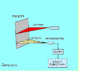
The image plate reader is another important component of the image acquisition control in computed radiography. The image reader converts the continuous analog information (latent image) on the imaging plate to a digital format. • In this unit the screen is scanned by a very small laser beam. When the laser beam strikes a spot on the screen it causes light to be produced (the stimulation process). The light that is produced is proportional to the x-ray exposure to that specific spot.
The result is that an image in the form of light is produced on the surface of the stimualible phosphor screen. A light detector measures the light and sends the data on to produce a digitized image. As the surface of the stimualible phosphor screen is scanned by the laser beam, the analog data representing the brightness of the light at each point is converted into digital values for each pixel and stored in the computer memory as a digital image.
Radiographic Condition
Computed radiography can be carried out under the same condition for general radiography by only replacing a conventional film / screen system with an imaging plate, dose and quality of X-ray, among the parameter given to images, are qualively different from those for screen-film system.
Gradian processing is done via computer operation to optimize image contras and optical density. Image contrast can be adjusted as desired, in accordance with the anatomical region and diagnostic purpose. The shape of gradian curve can also be changed, dark and white reversal can be easily archieved. Spatial Frequency is used for enhancing both the edges of anatomical region and structure of a certain size, and the basic contras curve is used as base from which contrast and density can be adjusted as fairly as desired. Image processing improves diagnostic accuracy and expand diagnostic scope.
Two types of image processing are involved : 1). Gradation Processing ; Incline of the gradation curve, shape of the gradation curve, density which determines incline of gradation curve, and degree of parallel movement of gradation curve. 2). Spatial Frequency Response Processing ; Frequency to emphasize in frequency processing, shape of emphasize curve depending on density frequency processing, Degree of emphasis in frequency processing.
Gradation Processing and Spatial Frequency Response Processing are use to control the contrast and density of the displayed image. Gradation Processing controls the range of densities use to display structures on the image ; Gradation Processing is similar to window setting used in CT-Scan. Spatial Frequency Response Processing controls the sharpness of boundaries betwen two structures of different density.
The wide dynamic range and linear response of the typical digital receptor is like a "two-edged sword". The advantage is that a wide range of exposures, and exposure errors, will still produce good image contrast. That is, the loss of contrast with exposure error is not a limiting factor as it is with film. Even though images with good contrast can be produced with relatively low exposures, they will have a high level of quantum noise. We recall from other modules that the level of image (quantum) noise depends on the exposure to the receptor. When a low exposure is used, the result can be excessive image noise. The other problem is that excessively high and unnecessary exposures can be used to form images. While these images will have good quality (low noise) there will be unnecessary exposure to the patient. This problem does not exist with film radiography because the increased exposure will result in a visibly overexposed film.
In general, for a radiographic procedure there is an optimum exposure that produces a good balance between image noise and patient exposure. The challenge to the technologist is to make sure that the technique factors are set to produce this optimum exposure.
Quality Characteristics
Computed radiography have the five specific quality characteristics : contrast, detail, spatial, artifacts and noise. The contrast sensitivity of a computed radiographic procedure and the image contrast depend on several factors. Two of these, the x-ray beam spectrum and the effects of scattered radiation are similar to film radiography. Computed radiography is the ability to adjust and optimize the contrast after the image has been recorded. This usually occurs through the digital processing of the image and then the adjustment of the window when the image is being viewed.
Detail is reduced and limited by the blurring that occurs at different stages of the imaging process both computed and film radiography are three sources of blurring:
• The focal spot (depends on size and object location)
• Motion (if it is present)
• The receptor (generally because of light spreading within the fluorescent or phosphor screen)
Computed radiography is that additional blurring is introduced by dividing the image into pixels. Each pixel is actually a blur. The size of a pixel (amount of blurring) is the ratio of the field of view (image size relative to the anatomy) and the matrix size. Pixel size is a factor that must be considered because it limits detail in the images.
The most predominant source of noise in digital radiography is generally the quantum noise associated with the random distribution of the x-ray photons received by the image receptor. The level of noise depends on the amount of receptor exposure used to produce an image. With computed radiography it can be adjusted over a rather wide range because of the wide dynamic range of the typical digital receptor. The noise is controlled by using the appropriate exposure factors.
Three factors are directly responsible for computed radiography image resolution : 1). The dimension of the crystals in the imaging plate, 2). The size of the laser beam in the reader, and 3). The image-reading matrix. Computed radiography contrast resolution is currently greater than that of conventional film, but spatial resolution is slightly less than that film.
Benefits of Computed Radiography
When using a computed radiography system certain benefits are readily apparent, including the following :
Improved Diagnostic Accuracy and Expanded Diagnostic Scope ; By storing laser-scanned x-ray images on high-sensitivity imaging plates, minute differences in x-ray absorbtion, are detected, providing highly detailed and easily readable diagnostic information. The wide exposure latitude permits diagnosis on an entire area of interest, allowing imaging from bones to soft tissue with a single exposure. Computer analysis of images can provide increased diagnostic information to assist in the medical treatment of a patient.
X-ray Dosage Reduction ; The high-speed imaging plate coupled with the efficient information read-out of the high-precision laser spot scanning device allows the patient to be exposed to a lower x-ray dose than that using a conventional film-screen system. This is especially worthwhile for pediatric examinations. Reductions vary somewhat with the type of examination performed, as shown in table below :
Comparison of radiographic film H and D response curve
To linear imaging plate response
Repeat Rate Reduction ; Because of the computed radiography system’s wide technique latitude, technical errors are easily corrected to provide prime diagnostic information. When a film-screen combination is used, technical errors in either direction can markedly degrade the image quality. Technical errors have much less effect on the final quality of a computed radiographic image. This benefit obviously increases throughput and reduces the patient’s discomfort, because it lessens the need to repeat the examination. The technical latitude of computed radiography is a tremendous asset in the area of portable radiography.
Teleradiographic Transmission ; Image Plate reader devices can be linked via dedicated phone lines, microwave transmission or other teleradiographic means to centralize the review of image data. This means of image sharing could obviously benefit affiliated hospitals or clinic that are separated by large geographic distances and share professional staff. Teleradiology could also provide immediate consultation with specialists, which benefits not only the patient but also the level of efficiency of the institution.
Department Efficiency ; The computed radiography system eliminates all darkroom work. With this factor plus the previous benefits, departemental efficiency is ultimately increased.
Summary
Computed Radiography uses image receptors with barium fluorohalide screens that function quite differently from intensifying screens in that a latent image is stored upon exposure to ionizing radiation. The latent image can be released upon stimulation by light. The incident photon beam produces a latent image within the fluorohalides. The flourohalides are stimulated by a laser, which causes them to emit light. The light beam is then detected by a photomultiplier tube which digitizes the image. Display and storage then proceed as with any other digital image. Although resolution continues to be a problem, pasien dose is dramatically reduced, and digital contrast and latitude control are superior.
THE BASIC PRINCIPLES OF COMPUTED RADIOGRAPHY
Labels:
Computed radiography (CR)
Subscribe to:
Post Comments (Atom)


3 comments:
Thanks for posting a wonderful information about CR system. I have read many articles about CR systems, but never got excellent information like this .
Useful Information provided!. Thanks for mentioning the features, applications, and other detailed information of the CR system. Hospital product Directory, Leading B2B Kindly visit our website. CR System
thanks for this exclusive information on CR.
Post a Comment